Sunday, January 5, 2014

Carbon Monoxide

Given the Planet Hoth like winter weather out there, we are likely to see a few cases of….

Carbon Monoxide Poisoning

Carbon Monoxide is a colorless, odorless and nonirritating gas produced by the incomplete combustion of carbonaceous materials. Thus, we often see an increase in presentations during the winter, secondary to the use of indoor heating sources. It is one of the most common causes of unintentional poisoning deaths in the United States.

Carbon Monoxide impairs oxygen delivery to the tissues because of the high binding of CO to hemoglobin. 240x the affinity of oxygen! And thus causes a left shift in the oxyhemoglobin dissociation curve.

High Risk Exposures: smoke inhalation, stoves, portable heaters, gasoline powder generators, automobile exhaust, charcoal grills, inhaling spray paint

In acute poisonings, you can see:
  • Central Nervous System: headache (most common symptom), malaise, confusion, depression, impulsiveness, hallucinations, agitation, drowsiness, visual disturbances, syncope, seizure, coma
  • Gastrointestinal symptoms: abdominal pain, nausea, vomiting, diarrhea
  • Respiratory symptoms: shortness of breath
  • Cardiac: chest pain, palpitations, myocardial injury
  • Delayed Neuropsychiatric syndrome: can arise 3 –240 days after recovery
  • Note: often diagnosed as a viral syndrome

Examination and Tests:
  • Suspect it! If you do not consider it, you cannot diagnosis it
  • Base work-up on the patient’s symptoms and clinical exam
  • HbCO … elevated levels are significant, but, a normal level does not rule out exposure
    • Level does not correlate well with the clinical signs and symptoms
    • Normal: 0.4-3%
    • Smokers: 0-5% (some can have up to 10-15%)
    • Mild poisoning: greater than 10% without clinical symptoms
    • Moderate poisoning: greater than 10% with minor clinical signs/symptoms (headache, fatigue)
    • Severe poisoning: greater than 20% with loss of consciousness, confusions or signs for cardiac ischemia
  • SpO2 is not helpful as standard pulse oximetry cannot screen for CO, as it does not differentiate carboxyhemoglobin from oxyhemoglobin
  • ABG
  • Lactic acid
  • BMET (look for hyperglycemia, hypokalemia, renal failure)
  • CPK (look for signs of rhabdomyolysis)
  • CXR (if there are pulmonary symptoms)
  • Head CT (in patients who do not improve rapidly)
  • ECG
  • Cardiac Enzymes (if abnormal ECG or history of cardiac disease)
  • Cyanide level (if smoke exposure)

Management:
  • Remove the patient from the source (duh!)
  • ABCs
  • Oxygen. Oxygen Administration enhances the elimination of carbon monoxide from the body.  If the patient is symptomatic in the Emergency Department, use a NRB.
    • Carbon Monoxide half-life on room air is 4-5 hours
    • Carbon Monoxide half-life on 100% NRB is about 60-90 minutes
    • Carbon Monoxide half-life with HBO treatment is about 30 minutes
    • Treat until asymptomatic
    • If pregnant, consult OB as they may need treatment for 24 hours
  • Consider transferring for hyperbaric treatment if COHb is greater than 25%, if there is evidence for ongoing end-organ ischemia, if there is severe metabolic acidosis (pH less than 7.1), if there was a loss of consciousness, or in pregnant women with a level greater than 20% or signs of fetal distress.
    • Note: These are controversial
  • Don't forget other potiental exposures!  Make sure everyone else has been removed from the environment

Thursday, January 2, 2014

Uncomplicated UTIs

Acute Uncomplicated Cystitis and Pyelonephritis

A common presentation to every Emergency Department, but practice variation is amazing.  What does the literature show?


Acute cystitis refers to an infection of the bladder (lower track) and it can occur alone or in conjunction with pyelonephritis (infection of the kidney, the upper track). 

We will focus on uncomplicated cases, in an otherwise healthy, non-pregnant adult. 

  • So, what makes it a complicated urinary tract infection?  Complicated UTIs are associated with underlying conditions that increase the risk of infection or of failing therapy (obstruction, anatomical abnormality, urologic dysfunction or multi-resistant uropathogens). 

The vast majority of uncomplicated cystitis and pyelonephritis in women are the result of E. coli (75-95%), with other common causes being Proteus mirabilis, Klebsiella penumoniae and Staph saprophyticus.  Therefore, your local antimicrobial susceptibility patterns to E. coli should be the basis for treatment (contact your ID colleagues and/or lab for this information).
 
Clinical manifestations of cystitis consist of dysuria, urinary frequency, urinary urgency, suprapubic pain, and/or hematuria (note: these can be subtle in the extremes of age).

Clinical manifestations of pyelonephritis consist of those of cystitis plus fever (>38oC), chills, flank pain, costrovertebral angle tenderness and nausea/vomiting. 

Diagnosis: on physical make note of temperature, costovertebral angle tenderness and an abdominal examination

Labs:
Urinalysis (either dipstick of microscopic) and urine culture with susceptibility
o   A urinalysis in the absence of a urine culture is sufficient for the diagnosis of uncomplicated cystitis if the symptoms are consistent with a UTI, unless there is reason to suspect antimicrobial resistance or other complicating features
  • Urinalysis: Pyuria (in a clean catch, mid-stream urine specimen) is the most valuable diagnostic test for UTI, as it is present in almost all women with acute cystitis or pyelonephritis; its absence prompts the evaluation for another diagnosis. 
o   An abnormal result is 10 leukocytes/mL
o   If white cell casts are noted, they are diagnostic of an upper tract infection. 
o   The presence of hematuria is helpful since it is common in UTI but not in urethritis or vaginitis. 
  • Urine Dipsticks look for the presence of:
o   Leukocyte esterase (an enzyme released by leukocytes, thus reflecting pyuria)
o   Nitrite (reflecting the presence of Enterobacteriaceae, which converts urinary nitrate to nitrite). 
o   The dipstick is most accurate for predicting UTI when it is positive for either leukocyte esterase or nitrite, with a sensitivity or 75% and a specificity of 82%
  • Urine culture. Routine cultures are not generally necessary.  Consider obtaining prior to initiation of therapy if: the symptoms are not characteristic of UTI, if the symptoms persist or if they have recur within 3 months following prior treatment or if a complicated UTI is suspected. 


Differential Diagnosis: vaginitis, urethritis, structural uretheral abnormalities, painful bladder syndrome, pelvic inflammatory disease, nephrolithiasis
           
Treament:
  • Cystitis:
·      Nitrofurantoin monohydrate/macrocrystals: 100mg PO BID for 5 days
o   Avoid if there is a suspicion for pyelonephritis and it is in contraindicated when creatinine clearance if < 60mL / min
·      Trimethoprim-sulfamethoxazole: 1 double strength tablet (160/800) BID for 3 days         
o   Avoid if location resistance is >20% or if the patient has taken TMP-SMX in the preceding 3 months
·      Fosfomycin trometamol: 3g in a single dose.  (more expensive and slightly less effective)
·      Fluoroquinolones (ciprofloxacin, levofloxacin, ofloxacin) for 3 days
o   Should be reserved for other uses other than acute cystitis
·      Beta-Lactams (amoxicillin-clavulanate, cefpodoxime, cefdinir & cefaclor) for duration of 7 days are less effective than TMP-SMX or the fluoroquinolones
o   Cephalexin is not well studied
o   Generally have inferior efficacy and more adverse effects compared with other agents, so they should be used with caution for uncomplicated cystitis
  • Pyelonephritis
·      Urine culture should be obtained
·      Outpatient management is appropriate for patients with mild-moderate illness who can be stabilized with rehydration and have adequate follow-up
·      Fluoroquinolones are the only oral antimicrobials recommend for the outpatient empirical treatment of acute complicated pyelonephritis
o   Ciprofloxacin 500mg BID for 7 days or 1000mg ER for 7 days; consider an initial 400mg intravenous dose
o   Levofloxacin 750mg PO for 5-7 days
o   An initial dose of a long-acting parental antimicrobial (ceftriaxone 1g or an aminoglycoside) can be considered prior to starting oral treatment, especially if local resistance to fluoroquinolones is greater than 10%. 
o   If allergic to fluoroquinolones, give an initial dose of a long-acting parental antimicrobial (ceftriaxone 1g or an aminoglycoside) and then start TMP-SMX or an oral beta-lactam for 14 days
o   Other alternative is aztreonam 1g IV every 8-12 hours

What about men?
  • Asymptomatic bacteriuria and symptomatic urinary tract infection are much less common in men due to the longer urethral length, drier periurethral environment and antibacterial substances in prostatic fluid. 
  • Risk factors: insertive anal intercourse and lack of circumcision
  • Treatment is similar to the treatment in women, except, nitrofurantoin and beta-lactams should not be used (do not achieve reliable tissue concentrations and are less effective for occult prostatitis. 
o   A 7 day course of antibiotics are recommended


References:
International Clinical Practice Guidelines for the Treatment of Acute Uncomplicated Cystitis and Pyelonephritis in Women: A 2010 Update by the Infectious Diseases Society of America and the European Society for Microbiology and Infectious Disease.  Clinical Infectious Disease. 2011; 52(5):e103-e120


Up-To-Date

Friday, December 20, 2013

Wellens Syndrome

Ok, so not every post on EverydayEmergencyMedicine will focus on the basics, some will focus on the rare/odd/funny cases that we see everyday as well.


Case Presentation:
Sorry, the blog post title gives the case away….

Male in his early 50s presents for a sexual transmitted infection (STI) check, along with his new (and younger) significant other; he is currently asymptomatic.  During the history, patient admits to some shortness of breath and chest discomfort during a recent argument with his significant other, as well as, some shortness of breath with exertion (both walking and intercourse) in the past few days.  Patient, again, has no current complaints.   

The history prompted the patient to have an ECG preformed.  Which showed….


Because of these findings, cardiology was contacted and the patient was admitted.


What does this ECG show?                       Wellens Syndrome


Wellens Syndrome represents a characteristic T-wave change on electrocardiogram (ECG) that is present during pain-free periods in a patient with intermittent chest pain and symptoms consistent with unstable angina/ischemic chest pain. 

The findings of Wellens Syndrome suggest a high-degree stenosis of the proximal left anterior descending (LAD) coronary artery (i.e., the “Widow Maker”), that if not treated with intervention will lead to a massive anterior myocardial infarction and/or death within two weeks (mean time, 8.5 days) in 75% of patients (i.e., a “Widow Maker”).  Medical Management is ineffective.

Wellens syndrome is characterized by T-wave changes in the precordial leads, mainly V2 & V3 ± V4, during pain free periods, in a patient with a history of chest pain that is consistent with unstable angina/ischemic chest pain.  Type A (or Type I) (75-76% of cases) shows deeply inverted T-waves, while Type B (or Type II) (24-25% of cases) shows biphasic T-wave.  Type A is rarely missed, but Type B is often interpreted as “non-specific T wave changes” by the computer and clinicians. 

Type A (or Type I)


Type B (or Type II)


The diagnosis should be made by the history obtained from the patient and ECG only, as cardiac enzymes will be normal or only mildly elevated.  This also helps enforce the importance of serial ECGs and pain-free ECGs in patients with a history of chest pain that is concerning for unstable angina/ischemic chest pain.

These patients do not necessarily need immediate catheterization, but they need urgent catheterization.  These patients should not undergo a stress test, as it places them at risk for an acute anterior wall myocardial infarction.  Cardiac catheterization has demonstrated patients with Wellens syndrome to have a 50% or greater stenosis of the proximal LAD.


Case resolution:
This patient’s ECG showed Wellens Type B (or Type II) … biphasic T waves in leads V2 & V3 (and arguably in leads in V1 & V4 as well), the computer read it as “T wave abnormality, consider anterior ischemia.”  The patient was asymptomatic in the emergency department and had negative cardiac markers and was admitted to cardiology (the patient was reluctant to be admitted).  Cardiology did order a treadmill stress test for the morning, which the patient failed and thus went to cardiac catheritzation and a 95% proximal LAD lesion was found and two stents were placed. 

Had this been missed, the patient likely would have passed in bliss during intercourse with his younger significant other (personal commentary).


References:
Rosen’s 7th Edition

De Zwaan C, Bar FW, Wellens HJ. Charcteristic electrocardiographic pattern indicating a critical stenosis high in the left anterior descenting artery in patients admitted because of impedning myocardial infarction. Am Heart J. 1982;103:730-6.

J Emerg Trauma Shock. 2009 Sept  Dec; 2(3): 206-208.


Monday, December 16, 2013

Hypothermia

Hypothermia
This will focus on accidental hypothermia; there are numerous secondary causes that will not be the focus of this review. 

Although typically associated with regions of the world with severe winters, hypothermia is also seen in areas with warmer climates, as well as during summer months and in hospitalized patients.  Even with modern supportive care, mortality for patients with moderate to severe accidental hypothermia approaches 40%.

Body Temperature reflects the balance between heat production (cellular metabolism) and heat loss (evaporation, radiation, conduction & convection).  The normal human core temperature is 98.6±0.9oF (37±0.5oC).  The human body has limited physiological capacity to respond to cold environmental conditions (basically, peripheral vasoconstriction), thus our behavioral adaptations of clothing and shelter.  In response to a cold stress, the hypothalamus attempts to stimulate heat production through shivering and increased thyroid, catecholamine and adrenal activity.  There is also sympathetic mediated vasoconstriction of peripheral tissues. 


Hypothermia is defined as a core temperature below 95oF (35oC).
  •          Mild: 93.2 – 95oF (34-35oC)
  •           Moderate: 86 – 93.2oF (30-34oC)
  •           Severe:  less than 86oF (less than 30oC)

Mild hypothermia is characterized by tachypnea, tachycardia, initial hyperventilation, ataxia, dysarthria, impaired judgment, shivering and “cold diuresis.”  “Cold Diuresis” is renal-fluid wasting due to hypothermia-induced vasoconstriction and diminished release of anti-diuretic hormone.

Moderate hypothermia is characterized by a proportionate reduction in pulse rate and cardiac output, hypoventilation, central nervous system depression, hyporeflexia, decrease renal blood flow, and the loss of shivering.  Patients may begin to display paradoxical undressing.  Atrial fibrillation, junctional bradycardia and other cardiac arrhythmias may also occur.

Patients with severe hypothermia develop pulmonary edema, oliguria, areflexia, hypotension, bradycardia, coma, ventricular arrhythmias and asystole.

Risk Factors for hypothermia include: Age (infants and the elderly), Environmental (exposure, drowning, and an alpine environment), Poverty, Homeless, Drugs / Toxicology and Psychiatric disorders.

To measure the temperature in a hypothermic patient requires a low-reading thermometer.  Most standard thermometers only read down to 93oF (34oC).  If the patient is conscious, a rectal probe thermometer is practical (although, to be truly accurate, needs to be inserted 15cm).  If the patient is intubated, an esophageal probe is preferred and is most accurate (inserted into the lower 1/3 of the esophagus). 


What studies to obtain?
  •  Finger stick glucose!  Do not miss the patient who is hypothermic secondary to hypoglycemia.  Remember, if the patient does not have glucose, the body cannot generate heat to help rewarm itself.  Also, insulin release is decreased in hypothermia, so hyperglycemia is common.
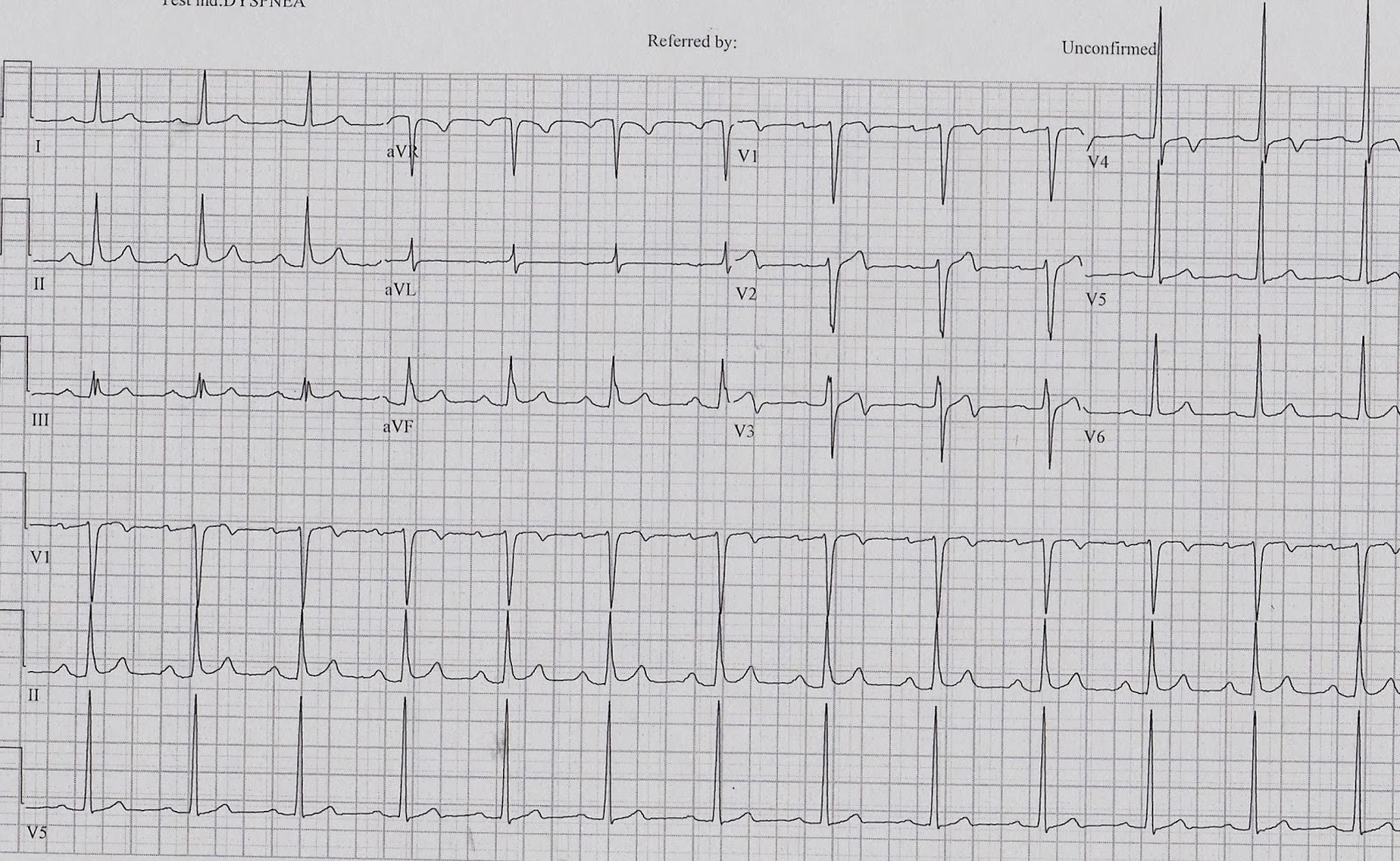
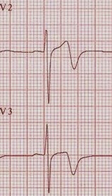
  • ECG
    •   Hypothermia causes characteristic ECG changes because of slowed impulse conduction through potassium channels.  This results in prolongation of all the ECG intervals.  There also maybe elevation of the J point, producing the characteristic Osborn J Wave (from the distortion of the earliest phase of membrane repolarization).                            
  • BMET
    • Resuscitation is futile if K >10
    • Check frequently during the resuscitation
  • CBC
    • There is 2% drop in Hct for each 1oC drop in temp
    • Thrombocytopenia is common
  • Lactate – elevated from shivering and poor tissue perfusion
  • PT/PTT – often coagulopathy is clinically evident but laboratory studies appear normal as the test is run at 98.6F (37C)
  •  Fibrinogen
  • Creatine phosphokinase
  • Arterial blood gas – acidosis often present due to severe respiratory depression and CO2 retention as well as lactic acid production
  • CXR – pneumonia (aspiration) is a common complication
  • Toxicology screen
  • ETOH

 
Management
  • ABCs
  • There is an alteration in ACLS algorithm for patients with severe hypothermia (less than 86oF (less than 30oC)).  In patients with severe hypothermia, begin CPR and attempt defibrillation once.  Withhold typical ACLS medications and any further defibrillation attempts until the patient’s core temp is >86oF (>30oC).  These patients will require active internal rewarming (information below).
  • Peripheral pulses may/will be difficult to assess, check a central pulse for up to a minute and consider using doppler.
  • Establish two large bore (14 or 16 gauge) peripheral intravenous lines and start an infusion of warmed (100.4 – 107.6oF (38 – 42oC)) isotonic crystalloid.  This will only really prevent further heat loss!  If central venous access is needed, use the femoral approach, if possible (to avoid the guide wire irritating the right atria and causing an arrhythmia, which can occur with the internal jugular or subclavian approach).
  • Treatment of cardiac arrhythmia.  Handle these patients with care!  Movement has been reported to trigger arrhythmias, including lethal ventricular fibrillation.  Remember, bradycardia is expected and pacing is not required unless the bradycardia persists despite rewarming to 90-95oF (32-35oC).
    • Typical progression is sinus bradycardia to atrial fibrillation to ventricular fibrillation to asystole.

  • Rewarming Therapies

o   Passive External Rewarming (PER).  This is the treatment of choice for patients with mild hypothermia.  Removal all wet and cold clothing and then cover the patient in blankets or other types of insulation (aluminum foil).  Set room temp to 82oF. PER requires the patient to have a physiologic reserve sufficient to generate heat by shivering and an increased metabolic rate.  If the patient’s temperature does not rise by 0.5-2oC/hr., reconsider the diagnosis (are they septic, hypoglycemic, hypovolemic, endocrine source, etc.) and consider starting AER.

o   Active External Rewarming (AER).  AER is indicated for moderate to severe hypothermia and for a patient with mild hypothermia who is unstable, lacks physiologic reserve or fails PER.  AER is a combination of warmed blankets, heating pads (watch for body surface burns from decreased sensation and reduced blood flow), radiant heat, warm baths, or forced warm air, which are applied to the patients skin. 
§  Core Temperature After Drop is a risk during AER.  This occurs when the truck and extremities are warmed simultaneously.  Cold, academic blood that has pooled in the extremities returns to the core and can cause a drop in temperature and pH.  This can trigger cardiac dysrhythmias. 
§  Rewarming shock can occur when peripheral vasodilation and venous pooling results in relative hypovolemia and hypotension.

o   Active Internal Rewarming is indicated for severe hypothermia and those who fail to respond to AER. 
§  Airway rewarming is utilized by use humidified air at 40-45oC. 
§  Pleural irrigation can be accomplished by placing two thoracotomy tubes (36 to 40 French), one placed anterior and one posterior, and instilling warmed IVF into the anterior chest tube and allowed to drain out the posterior chest tube.  If the patient is pulseless, use the left thoracic (bath the heart in the warm fluid), if the patient has a pulse, use the right thoracic to avoid triggering an arrhythmia by irritating the heart with tube insertion.
§  Bladder irrigation is another option

o   Extreme options (not readily available in many EDs): ECMO, hemodialysis, cardiopulmonary bypass

  • Medications

o   Glucose, if the patient is hypoglycemic
o   Naloxone
o   Thiamine, as patients are often alcoholics (also, Wernicke’s)
o   Hydrocortisone, if the patient has a history of adrenal insufficiency
o   Antibiotics, for suspected sepsis


Remember, the patient is not dead until they are “warm and dead.”  But, how warm is warm?  Target a temperature of 89.6oF (32oC) in adults and a temperature of 95oF (35oC) in children. 

If the body is frozen and chest compressions are impossible; or if the nose and mouth are blocked by ice; or if the patient’s potassium is >10, then resuscitation can be withheld. 


References:
Tintinalli Sixth Edition
Accidental hypothermia in Adults. Up-To-Date
Circulation. 2005;112:IV-136-IV-138.
Brown, et al. Accidental Hypothermia. N Engl J Med 2012;367:1930-8.